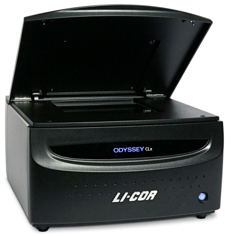
The Odyssey CLx is the next generation multifunctional imaging platform that can provide a wide range of applications. With Odyssey CLx, you can
- Get consistent, reproducible digital images, without the hassles and unpredictability of film.
- Analyze your data right away and plan your next experiment. – It’s already in digital format.
- One digital file contains all your image data. See both strong and faint bands clearly in the same image, with great sensitivity and no image saturation.
- Multiplex with two fluorescent colors to see even more data. Capture all the detail and complexity of your data.
- Near-infrared (NIR) fluorescence delivers consistent signals that aren’t affected by timing.
Select the right IRDye® or VRDye™ secondary antibody for your application using information in the table below.

Here are some general guidelines to help you choose the right secondary antibodies:
- Choose IRDye 800CW secondary antibodies to get low background and high sensitivity in the 800nm channel. Use IRDye 800CW for targets where your require the highest sensitivity. Dilution working range 1:10,000 – 1:40,000.
- Choose IRDye 680RD secondary antibodies to get low background and for general use when detecting in the 700nm channel. Start using IRDye 680RD first over other 700 nm dyes. Dilution working range 1:10,000 – 1:40,000.
- Choose IRDye 680LT secondary antibodies to get high signal and for specific uses of detection in the 700nm channel. These antibodies are not recommended when getting up and running on system. Once established near-infrared protocols are optimized with IRDye 680RD, IRDye 680LT can be used to optimize signals in the 700 channel. Dilution range 1:20,000 – 1:40,000. Note: optimization may be required with IRDye 680LT. Please see pack insert for specific detergents/protocol modifications needed.
Odyssey CLx Applications:
- Western blots: Two-Color Infrared and In-Gel
- Cell-Based Assays: In-Cell Western™ and On-Cell Western
- Protein Detection: Coomassie-Stained Gels, Membrane and Slide Arrays
- Small Animal Imaging: In Vivo, Whole Organ, and Tissue Section
- Nucleic Acid Detection: Mobility Shift Assays,
- DNA Gel Staining (Syto®60), and Arrays
- Microwell Assays: ELISA/FLISA, Protein Arrays, and RNAi Analysis